No active uploads
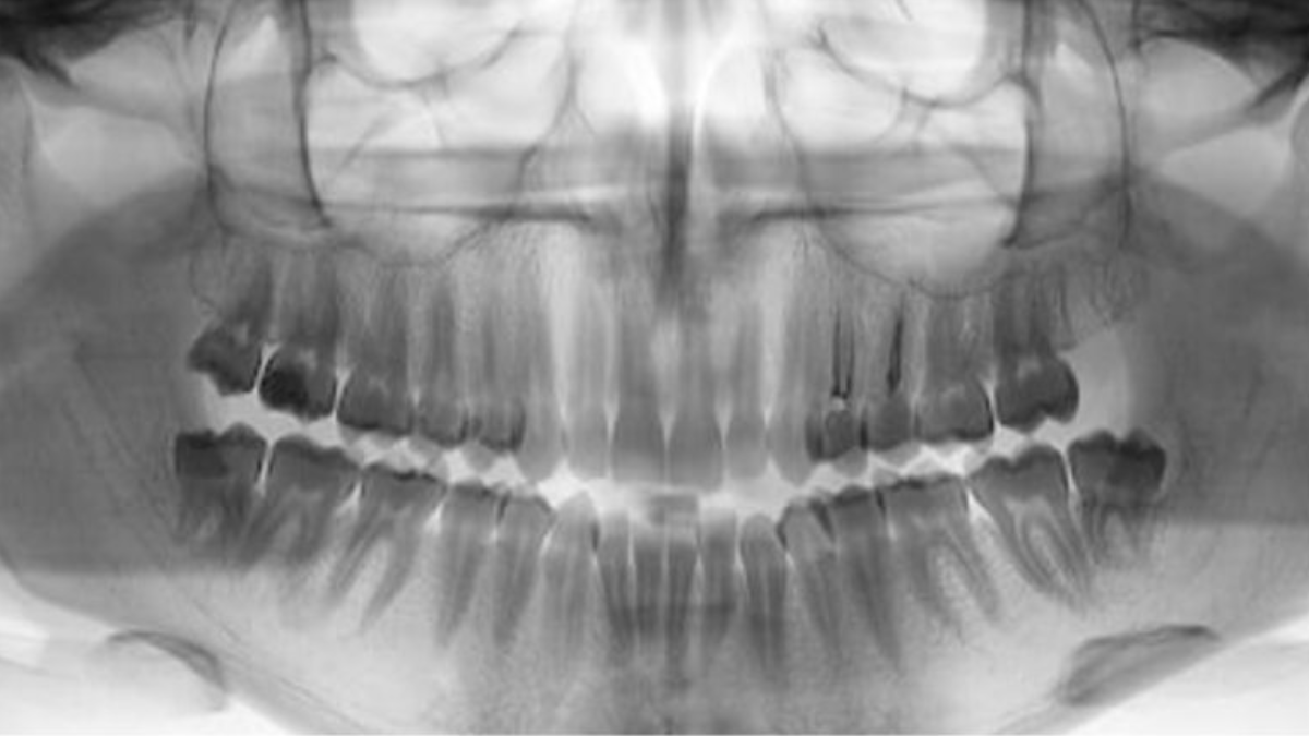

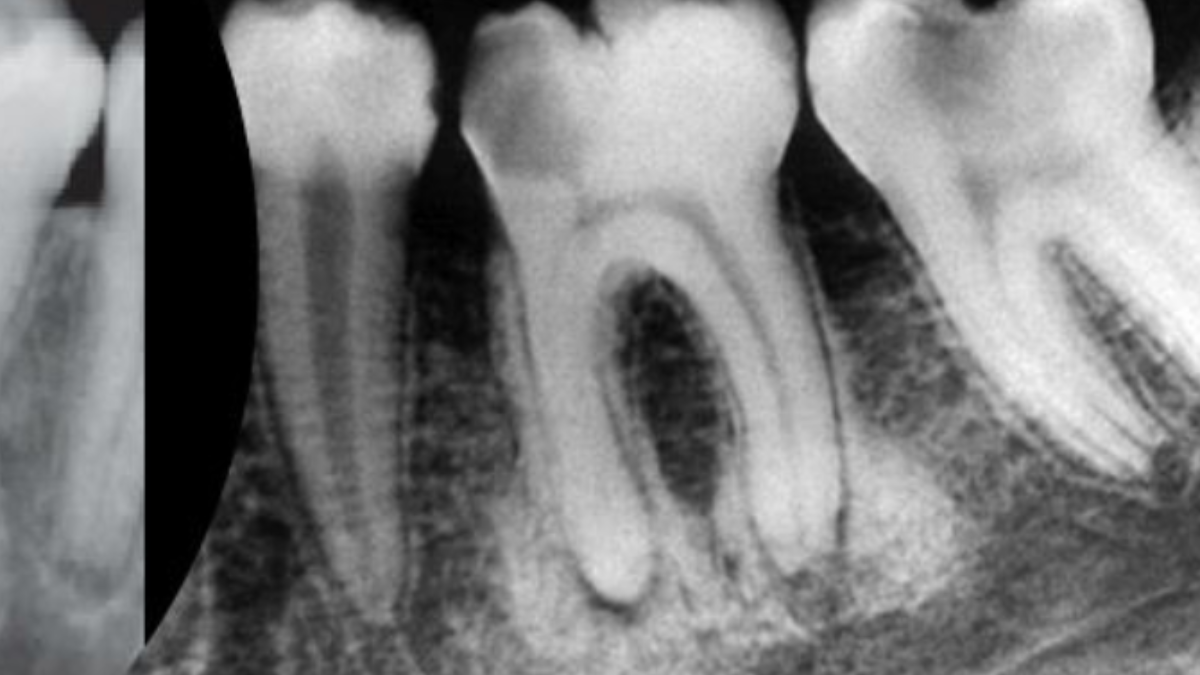
Dental professionals must understand the difference between normal anatomical landmarks and abnormal findings, such as artifacts or pathology, which may be present on a panoramic image when viewing both the mandible and maxilla in the one projection. It is recommended to review the image systematically in order not to overlook anything that might be a deviation from normal. The focal trough is the area in which structures will appear most sharply and clearly. Structures, which fall in front of or behind, the focal trough, can be distorted, magnified or reduced. Panoramic radiography offers several advantages over conventional intraoral radiography. Broad anatomic region is imaged, including additional visualization of the areas of the body of the mandible beyond the periapical region, the ramus, the temporomandibular joint, the maxillary sinus and the stylohyoid complex. The focal trough is the area in which structures will appear most sharply and clearly. Magnification, geometric distortion, and overlapped images of teeth sometimes occur. Objects situated outside the focal trough will be distorted or obscured on the radiograph.
Dentoflow nevertheless undertakes to:
- Act promptly to remove any illicit or inappropriate content as soon as it becomes aware of it
- Provide users with a feature allowing them to contact the company organizing the event, accessible via the event summary in the "Events" section of the user account.
- Set up an effective reporting process for users wishing to report a problem related to an event or a company.
Participants are encouraged to address any questions or complaints directly to the event organizing company.